
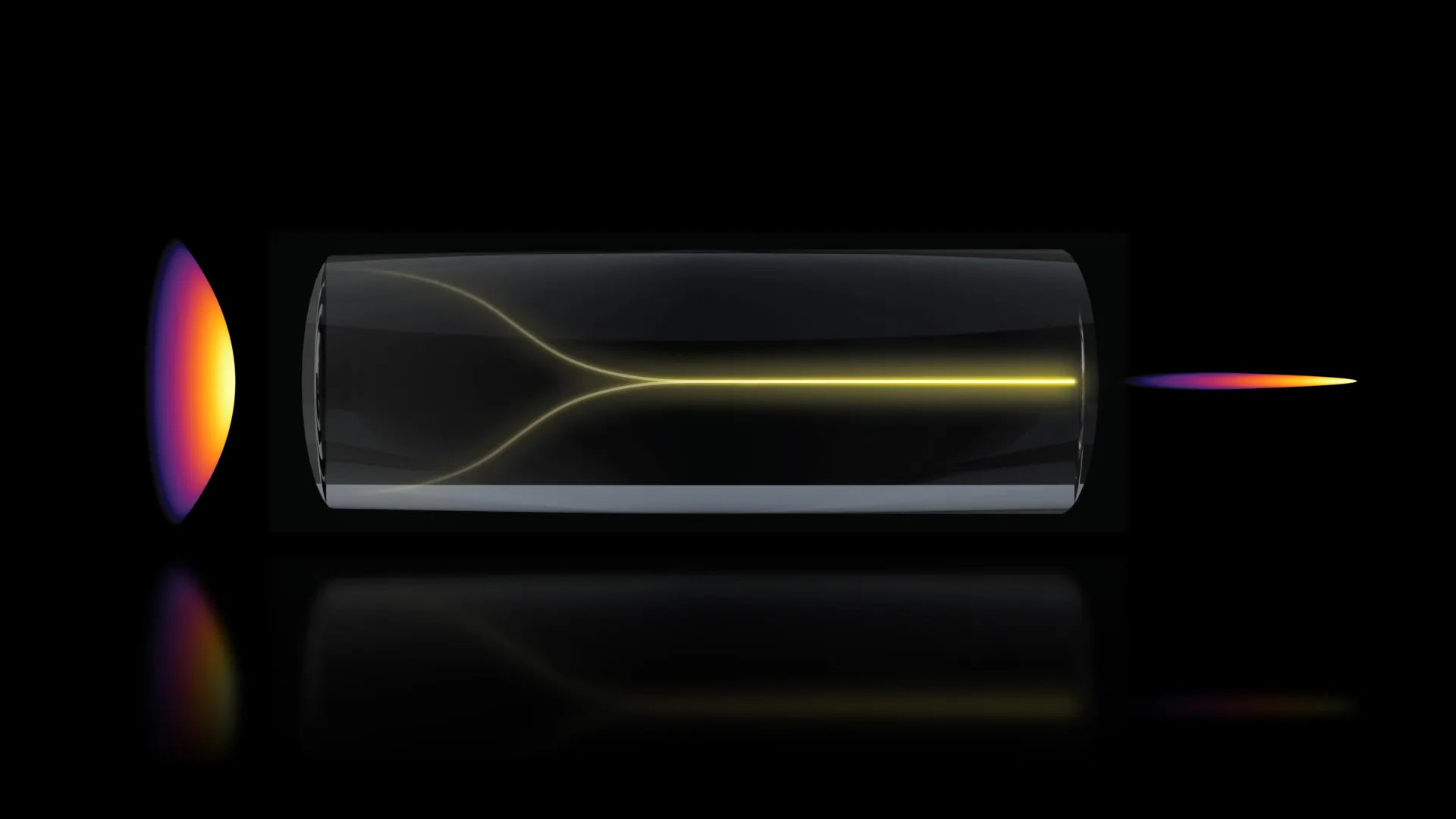
Researchers at MIT have identified an unexpected effect in optical physics that could lead to a faster and more detailed way to image living tissue. Under specific conditions, what normally looks like a scattered and disordered laser signal can reorganize itself into a narrow, highly focused “pencil beam.”
With this self-formed beam, the team produced 3D images of the human blood-brain barrier at speeds about 25 times faster than the current gold-standard approach, while preserving similar image quality. The method also makes it possible to watch individual cells absorb drugs in real time. This could help scientists evaluate whether treatments for conditions such as Alzheimer’s or ALS are actually reaching their intended targets in the brain.
“The common belief in the field is that if you crank up the power in this type of laser, the light will inevitably become chaotic. But we proved that this is not the case. We followed the evidence, embraced the uncertainty, and found a way to let the light organize itself into a novel solution for bioimaging,” says Sixian You, assistant professor in the MIT Department of Electrical Engineering and Computer Science (EECS), a member of the Research Laboratory for Electronics, and senior author of a paper on this imaging technique.
She is joined on the paper by lead author Honghao Cao, an EECS graduate student; EECS graduate students Li-Yu Yu and Kunzan Liu; postdocs Sarah Spitz, Francesca Michela Pramotton, and Federico Presutti; Zhengyu Zhang PhD ’24; Subhash Kulkarni, an assistant professor at Harvard University and the Beth Israel Deaconess Medical Center; and Roger Kamm, the Cecil and Ida Green Distinguished Professor of Biological and Mechanical Engineering at MIT. The paper appears today in Nature Methods.
A Surprising Laser Behavior Emerges
The finding began with an observation that did not fit expectations.
The researchers had previously built a precise fiber shaper, a device that allows careful control of laser light traveling through a multimode optical fiber, which is capable of carrying high levels of power.
Cao gradually increased the laser power to test the limits of the fiber.
Normally, increasing power causes the light to scatter more due to imperfections inside the fiber. Instead, as the power approached the threshold where the fiber might be damaged, the light suddenly concentrated into a single, extremely sharp beam.
“Disorder is intrinsic to these fibers. The light engineering you typically need to do to overcome that disorder, especially at high power, is a longstanding hassle. But with this self-organization, you can get a stable, ultrafast pencil beam without the need for custom beam-shaping components,” You says.
Conditions That Enable Self-Organizing Light
To reproduce this effect, the team identified two key requirements.
First, the laser must enter the fiber at a perfectly aligned, zero-degree angle, which is stricter than standard practice. Second, the power must be increased until the light begins interacting directly with the glass material of the fiber.
“At this critical power, the nonlinearity can counter the intrinsic disorder, creating a balance that transforms the input beam into a self-organized pencil beam,” Cao explains.
Such conditions are rarely explored because researchers typically avoid high power levels to prevent damaging the fiber. Precise alignment is also not usually necessary since multimode fibers can already carry large amounts of energy.
When combined, however, these factors allow the system to produce a stable beam without complex optical engineering.
“That is the charm of this method — you could do this with a normal, optical setup and without much domain expertise,” You says.
Sharper Imaging With Fewer Artifacts
Tests showed that this pencil beam is both stable and highly detailed compared to similar beams. Many conventional beams produce “sidelobes” — blurred halos that reduce image clarity.
In contrast, this beam remains clean and tightly focused.
The researchers then applied the technique to image the human blood-brain barrier, a dense layer of cells that shields the brain from harmful substances but also blocks many drugs.
Faster 3D Imaging of the Blood-Brain Barrier
Scientists often need to observe how drugs move through the blood vessels in this barrier and whether they successfully reach brain tissue. Traditional optical methods typically capture one 2D slice at a time, requiring repeated scans to build a complete 3D image.
Using the new pencil beam approach, the team generated rapid, high-precision images while also tracking how cells absorb proteins in real time.
“The pharmaceutical industry is especially interested in using human-based models to screen for drugs that effectively cross the barrier, as animal models often fail to predict what happens in humans. That this new method doesn’t require the cells to have a fluorescent tag is a game-changer. For the first time, we can now visualize the time-dependent entry of drugs into the brain and even identify the rate at which specific cell types internalize the drug,” says Kamm.
“Importantly, however, this approach is not limited to the blood-brain barrier but enables time-resolved tracking of diverse compounds and molecular targets across engineered tissue models, providing a powerful tool for biological engineering,” Spitz adds.
The system produced cellular-level 3D images with improved quality and did so roughly 25 times faster than existing methods.
“Usually, you have a tradeoff between image resolution and depth of focus — you can only probe so far at a time. But with our method, we can overcome this tradeoff by creating a pencil-beam with both high resolution and a large depth of focus,” You says.
Future Applications and Next Steps
Looking ahead, the researchers aim to better understand the physics behind this self-organizing beam and the mechanisms that allow it to form. They also plan to extend the method to other applications, including imaging neurons, and to explore ways to bring the technology into practical use.
This work was funded, in part, by MIT startup funds, the National Science Foundation (NSF), the Silicon Valley Community Foundation, Diacomp Foundation, the Harvard Digestive Disease Core, a MathWorks Fellowship, and the Claude E. Shannon Award.