

A new study in The Lancet Digital Health suggests the brain can respond to stroke in a surprising way. Researchers at the USC Mark and Mary Stevens Neuroimaging and Informatics Institute (Stevens INI) found that people with severe physical impairments after a stroke may show signs of a “younger” brain structure in areas that were not damaged. This appears to reflect how the brain adapts and reorganizes itself after injury.
The research was conducted as part of the Enhancing NeuroImaging Genetics through Meta-Analysis (ENIGMA) Stroke Recovery Working Group. Scientists analyzed brain scans from more than 500 stroke survivors collected across 34 research centers in eight countries. By applying deep learning models trained on tens of thousands of MRI scans, the team estimated the “brain age” of different regions in each hemisphere and examined how stroke affects both structure and recovery.
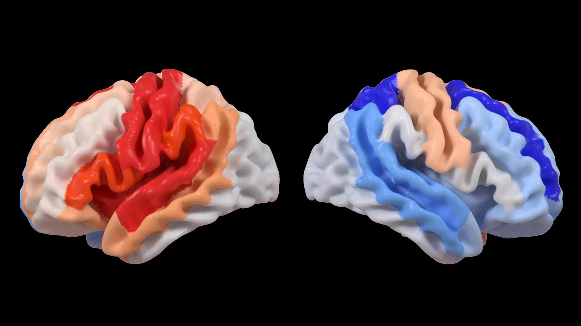
“We found that larger strokes accelerate aging in the damaged hemisphere but paradoxically make the opposite side of the brain appear younger,” said Hosung Kim, PhD, associate professor of research neurology at the Keck School of Medicine of USC and co-senior author of the study. “This pattern suggests the brain may be reorganizing itself, essentially rejuvenating undamaged networks to compensate for lost function.”
AI Reveals Brain Rewiring After Stroke
To carry out the analysis, researchers used a type of artificial intelligence called a graph convolutional network. This system estimated the biological age of 18 brain regions based on MRI data. They then compared this predicted age with each person’s actual age, a measure known as the brain-predicted age difference (brain-PAD), which serves as an indicator of brain health.
When these brain age measurements were compared with motor function scores, a clear pattern emerged. Stroke survivors with severe movement impairments, even after more than 6 months of rehabilitation, showed younger-than-expected brain age in regions opposite the site of injury. This effect was especially strong in the frontoparietal network, which plays an important role in movement planning, attention, and coordination.
“These findings suggest that when stroke damage leads to greater movement loss, undamaged regions on the opposite side of the brain may adapt to help compensate,” Kim explained. “We saw this in the contralesional frontoparietal network, which showed a more ‘youthful’ pattern and is known to support motor planning, attention, and coordination. Rather than indicating full recovery of movement, this pattern may reflect the brain’s attempt to adjust when the damaged motor system can no longer function normally. This gives us a new way to see neuroplasticity that traditional imaging could not capture.”
Large-Scale Data Reveals Hidden Patterns
The study relied on ENIGMA, a global collaboration that combines data from more than 50 countries to better understand the brain across different conditions. By standardizing MRI data and clinical information from many research groups, the team created the largest stroke neuroimaging dataset of its kind.
“By pooling data from hundreds of stroke survivors worldwide and applying cutting-edge AI, we can detect subtle patterns of brain reorganization that would be invisible in smaller studies. These findings of regionally differential brain aging in chronic stroke could eventually guide personalized rehabilitation strategies,” said Arthur W. Toga, PhD, director of the Stevens INI and Provost Professor at USC.
Toward Personalized Stroke Recovery
The researchers plan to continue this work by following patients over time, from the early stages after a stroke through long-term recovery. Tracking how brain aging patterns and structural changes evolve could help doctors tailor treatments to each person’s unique recovery process, with the goal of improving outcomes and quality of life.
Learn more about associations between contralesional neuroplasticity and motor impairment by viewing this video made by the Stevens INI.
The study, “Deep learning prediction of MRI-based regional brain age reveals contralesional neuroplasticity associated with severe motor impairment in chronic stroke: A worldwide ENIGMA study,” was funded by the National Institutes of Health (NIH) grant R01 NS115845 and supported by international collaborators from institutions including the University of British Columbia, Monash University, Emory University, and the University of Oslo.