

Researchers at Caltech and USC have created a new medical imaging approach that quickly produces 3D color images showing both the physical structure of soft tissue and how blood vessels are working. The technique has already been used to image several parts of the human body. Scientists say it could lead to better breast cancer imaging, improved tracking of nerve damage linked to diabetes, and new ways to study the brain.
Details of the work were published in Nature Biomedical Engineering.
Why Existing Imaging Tools Fall Short
Standard ultrasound is fast, affordable, and widely used, but it mainly shows tissue shape in two dimensions and offers a limited viewing area. Photoacoustic imaging provides a different kind of information. It works by sending laser light into the body and detecting the sound waves produced when certain molecules absorb that light. This allows doctors and researchers to see blood vessels in optical color and observe blood flow through arteries and veins. However, photoacoustic imaging does not capture detailed tissue structure well.
Other common imaging methods, including computed tomography (CT) and magnetic resonance imaging (MRI), come with tradeoffs. These techniques may require contrast agents, expose patients to ionizing radiation, cost more, or take too long to use frequently.
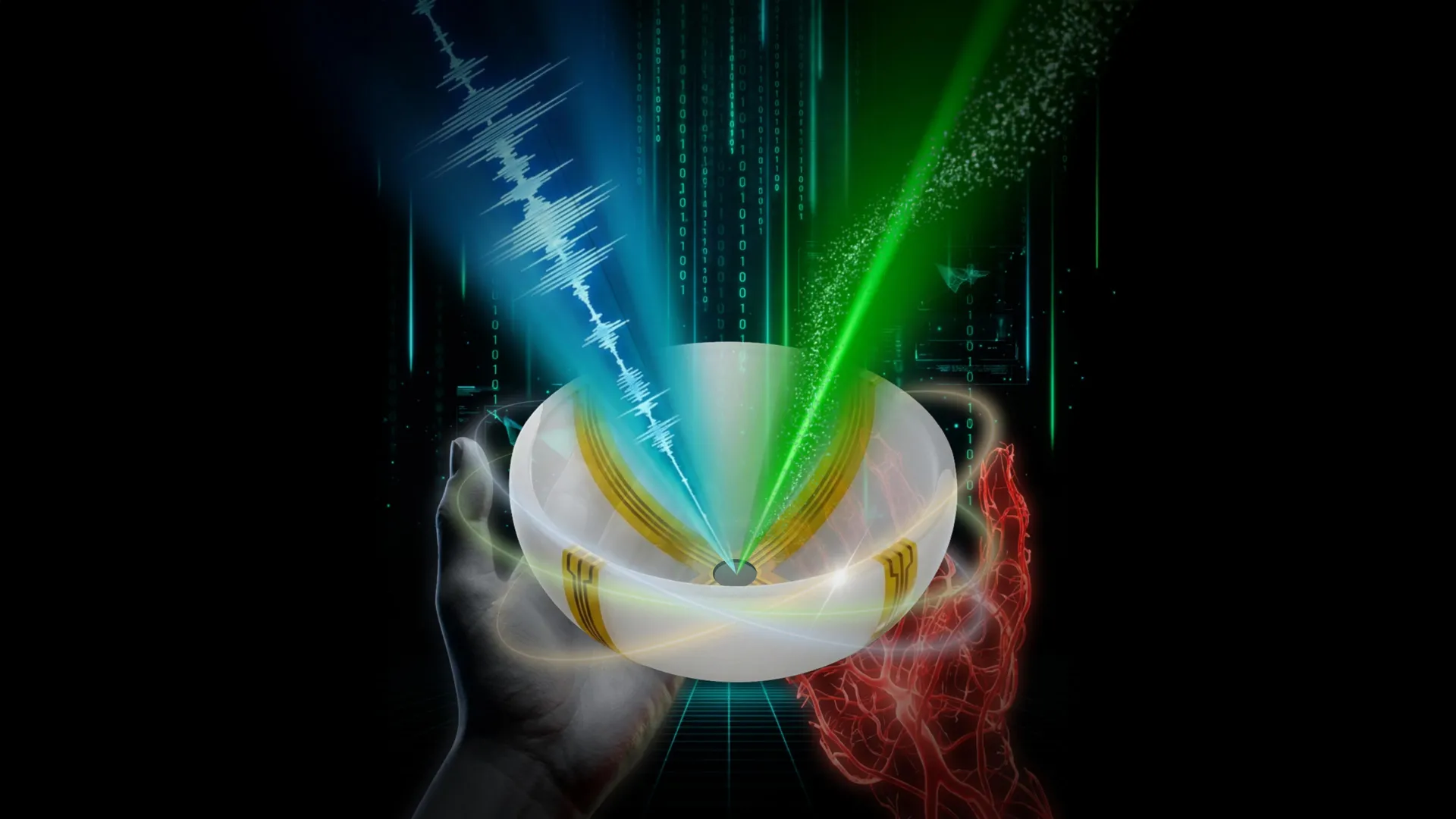
Combining Ultrasound and Photoacoustic Imaging
To overcome these limitations, the research team developed RUS-PAT (rotational ultrasound tomography, RUST, combined with photoacoustic tomography, PAT). Photoacoustic tomography was first developed more than two decades ago by Lihong Wang, the Bren Professor of Medical Engineering and Electrical Engineering and the Andrew and Peggy Cherng Medical Engineering Leadership Chair at Caltech. In PAT, tissue molecules that absorb light vibrate after being hit by short laser pulses, producing acoustic signals that can be measured and converted into detailed images.
Wang, who also serves as Caltech’s executive officer for medical engineering, said the goal of the new project was to merge the strengths of ultrasound and photoacoustic imaging. “But it’s not like one plus one,” he explains. “We needed to find an optimal way of combining the two technologies.”
A Simpler and More Practical Design
Traditional ultrasound systems rely on many transducers to send and receive sound waves, making direct integration with photoacoustic imaging too complicated and costly for broad use. Photoacoustic imaging, by contrast, only needs ultrasound detection. This difference led Wang to a new idea. “I thought, ‘Wait, can we just mimic light excitation of ultrasound waves in photoacoustic tomography, but do it ultrasonically?'”
In photoacoustic imaging, laser light spreads through tissue and triggers ultrasound waves that can be measured. Wang realized that a single wide-field ultrasound transducer could instead send sound waves throughout the tissue. The same detectors could then capture signals from both imaging methods.
The final system uses a small number of arc-shaped detectors that rotate around a central point. This setup effectively functions like a full hemispheric detector, while remaining far simpler and less expensive.
Demonstrated Potential for Human Use
“The novel combination of acoustic and photoacoustic techniques addresses many of the key limitations of widely used medical-imaging techniques in current clinical practice, and, importantly, the feasibility for human application has been demonstrated here in multiple contexts,” says Dr. Charles Y. Liu, a co-author of the study and a visiting associate in biology and biological engineering at Caltech. Liu is also a professor at the Keck School of Medicine of USC, director of USC’s Neurorestoration Center, and chair of neurosurgery at the Rancho Los Amigos National Rehabilitation Center.
Because the method can be used anywhere light can reach, RUS-PAT may have wide clinical applications. In breast cancer imaging, it could help doctors pinpoint a tumor’s location while also revealing information about its biological activity. For patients with diabetic neuropathy, the technique could allow physicians to monitor both nerve structure and oxygen supply in a single scan. Wang also notes its potential for brain research, where scientists could study brain anatomy while simultaneously observing blood flow dynamics.
Speed, Depth, and Early Testing
At present, the system can image tissue up to about 4 centimeters deep. Light can also be delivered using endoscopic tools, which may allow access to deeper areas of the body. Each RUS-PAT scan takes less than one minute.
The current setup places ultrasound transducers and a laser beneath a scanning bed. The system has already been tested on human volunteers and patients and is now in the early stages of moving toward clinical use.
Study Details and Funding
The paper is titled “Rotational ultrasound and photoacoustic tomography of the human body.” The co-lead authors are Yang Zhang, Shuai Na, and Dr. Jonathan J. Russin. Zhang and Na conducted the work as postdoctoral researchers at Caltech and are now based at Tsinghua University and Peking University in Beijing, respectively. Russin is affiliated with the Keck School of Medicine of USC and the Rancho Los Amigos National Rehabilitation Center in Downey, California.
Additional Caltech contributors include Karteekeya Sastry, Li Lin (PhD ’20), Junfu Zheng, Yilin Luo, Xin Tong (MS ’21), Yujin An, Peng Hu (PhD ’23), and former research scientist Konstantin Maslov. Lin is currently at Zhejiang University in Hangzhou, China. Dr. Tze-Woei Tan from the Keck School of Medicine of USC is also a co-author. The research was funded by the National Institutes of Health.